10801 West 87th Street, Overland Park, KS 66214

ADVANCING THE ART AND SCIENCE OF COSMETIC AND IMPLANT DENTISTRY

When needed, our prosthodontist can provide treatment for impacted teeth in Overland Park, Kansas. Please contact Reconstructive and Implant Dental Center at 913-534-8801 today to learn more and to schedule a visit with Dr. EDward M. Amet.

The Piezosurgery® Dental unit is designed for a wide variety of applications, including sinus lift procedures, extractions, bone block harvesting, bone graft harvesting, bone contouring, endodontic surgery, orthodontic micro surgery, ridge expansion, crown lengthening, periodontal therapy, implant site preparation and tori removal.

The Piezosurgery Dental unit uses high-frequency ultrasonic vibrations (24.7-29.5 kHz) to cut osseous tissue while safely avoiding damage to soft tissues. Sterile irrigating fluids are delivered through either a sterile fluid bag or bottle. All parts of the unit through which fluids flow, including the handpiece cords and handpiece, are sterilizable. This allows delivery of sterile irrigation while eliminating labor-intensive manual irrigation procedures.

The Piezosurgery Dental unit has one handpiece attachment. The unit is operated using an interactive keyboard with a digital display and multiple programmable settings. A foot pedal control is also available. The settings include four different power level options based on bone quality. Electrical requirements are 110VAC 10%, 60 Hz.

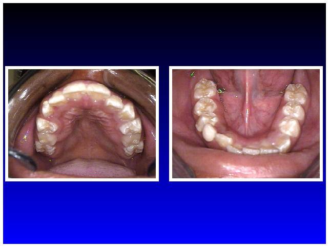
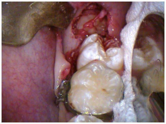
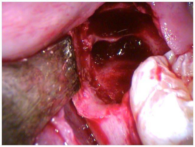
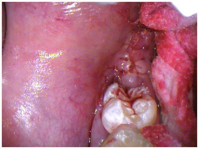
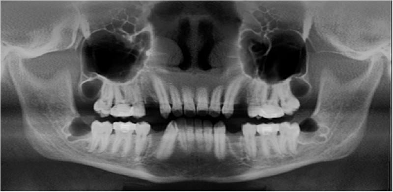
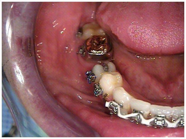
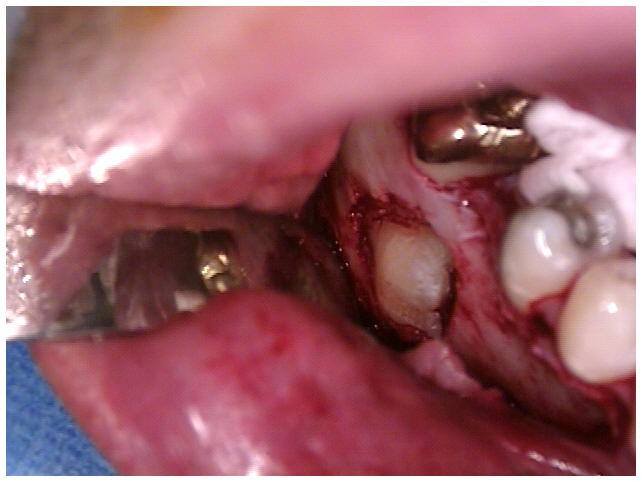
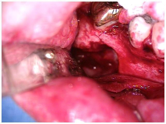
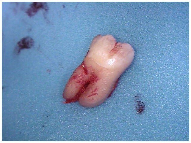
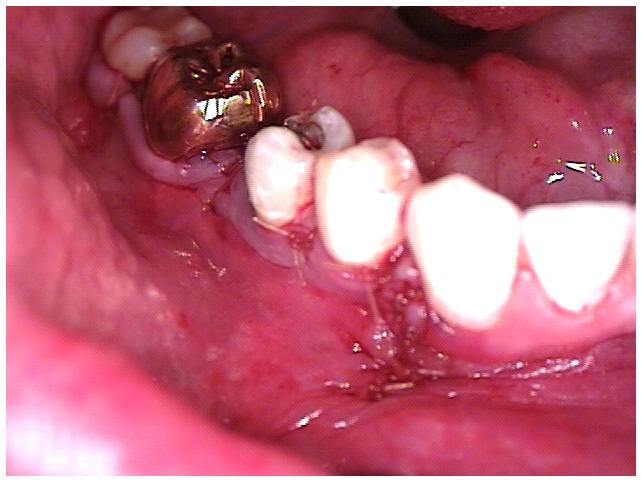
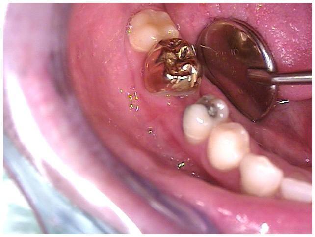
Case 1: Piezosurgery® – Horizontally Impacted First Molars

Pre-Operative Photograph

Fig 1. Pre-Operative Photograph

 Pre-Operative Radiograph

Fig 2. Pre-Operative Radiograph

Full Thickness Mucoperiosteal

Fig 3. Full Thickness Mucoperiosteal Flap, Removal of Buccal Bone, Piezosurgery


Removal of Horizontally

Fig 4. Removal of Horizontally Impacted First Molar

Horizontally Impacted

Fig. 5 Horizontally Impacted First Molar Rotated Out

 Horizontally Impacted

Fig 6. Horizontally Impacted First Molar Rotated Out With Sectioned Crown


 Horizontally Impacted

Fig 7. Horizontally Impacted First Molar Rotated out With Sections of Crown

Irradiated Cancellous

Fig 8. Irradiated Cancellous Bone Graft

Tissue Closure

Fig 9. Tissue Closure With 4-0 Gut Sutures Day of Surgery


Tissue Closure (1)

Fig 10. Tissue Closure 3 Days Post-Op Surgery

Third Day (2)

Fig 10. Third Day Post-Operative Radiograph

Tissue Closure

Fig 11. Tissue Closure 14 Days Post-Op Surgery


Tissue Closure

Fig 12. Tissue Closure 14 Days Post-Op Surgery


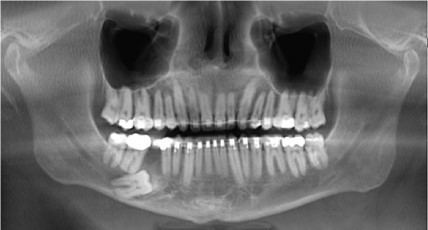
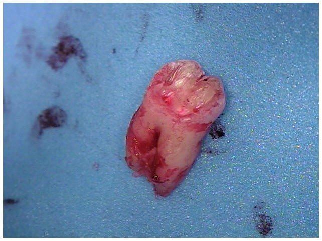
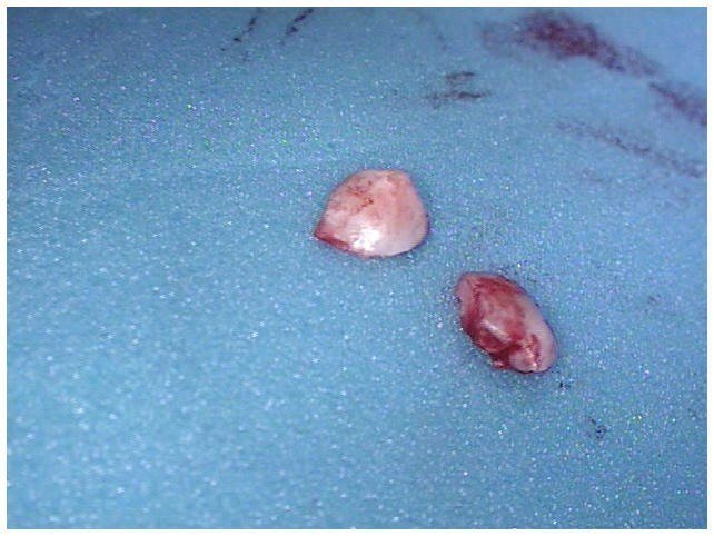
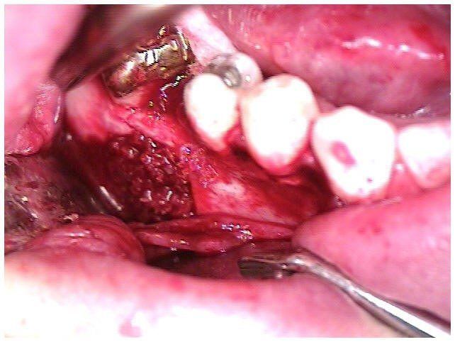
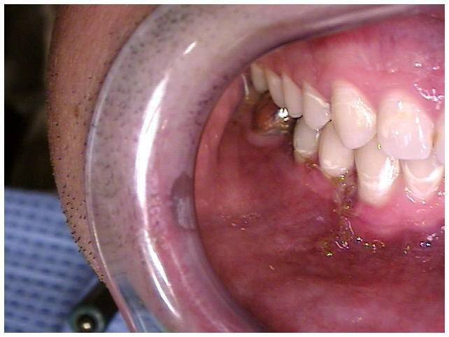
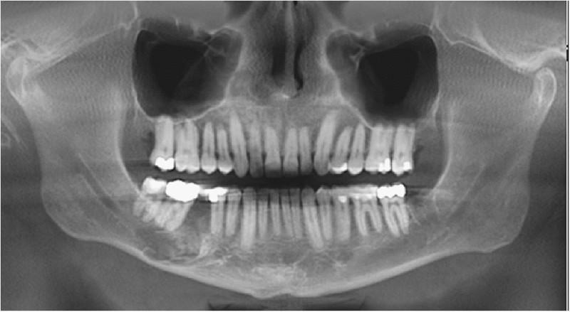
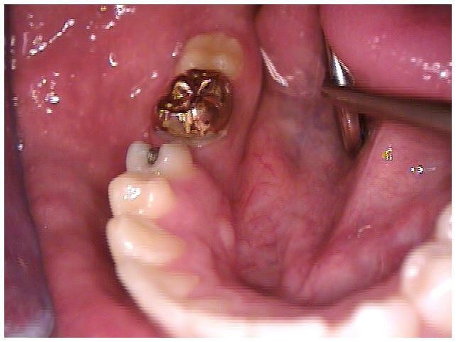
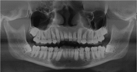
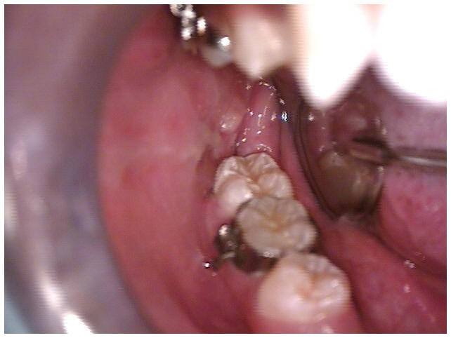
Case 2: Piezosurgery – Vertically & Horizontally Impacted Third Molars

Pre-Operative Photograph

Fig 1. Pre-Operative Photograph

Pre-Operative Radiograph

Fig 2. Pre-Operative Radiograph

Full Thickness

Fig 3. Full Thickness Mucoperiosteal Flap, Removal of Bone, Piezosurgery


Removal of Vertically

Fig 4. Removal of Vertically Impacted Third Molar

 Tissue Closure

Fig 5. Tissue Closure With 4-0 Gut Sutures Day of Surgery

Tissue Closure

Fig 6. Tissue Closure 7 Days Post-Op Surgery


 Third Day

Fig 7. Third Day Post-Operative Radiograph