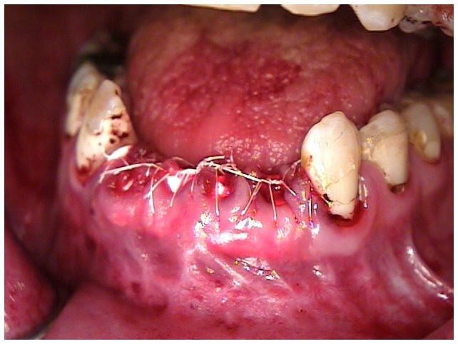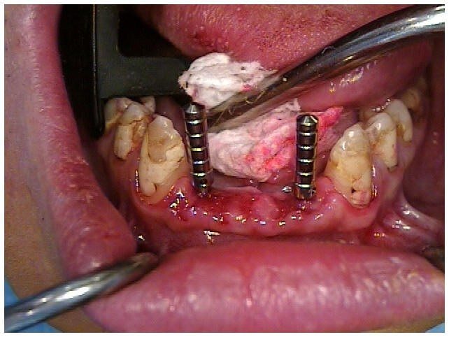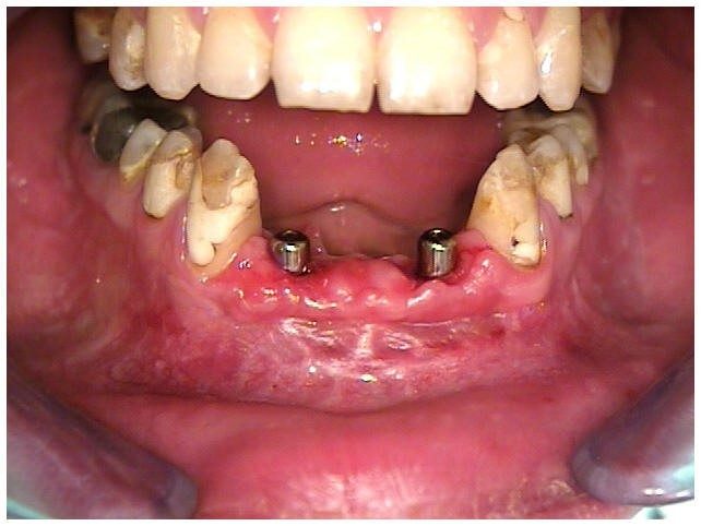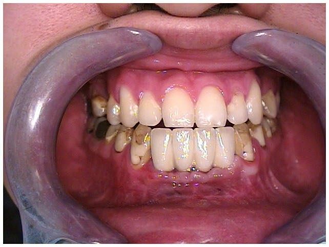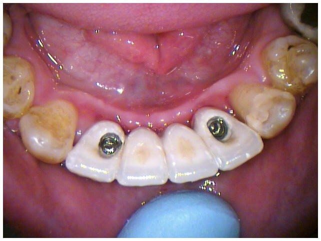
Successful implant treatment is directly related to achieving integration and restoring hard and soft supporting structures, aesthetics and function. It is necessary for the clinician to visualize the final prosthetic result before implant placement and to have a thorough understanding of the surgical and prosthodontic phases of treatment to achieve a predictable outcome.
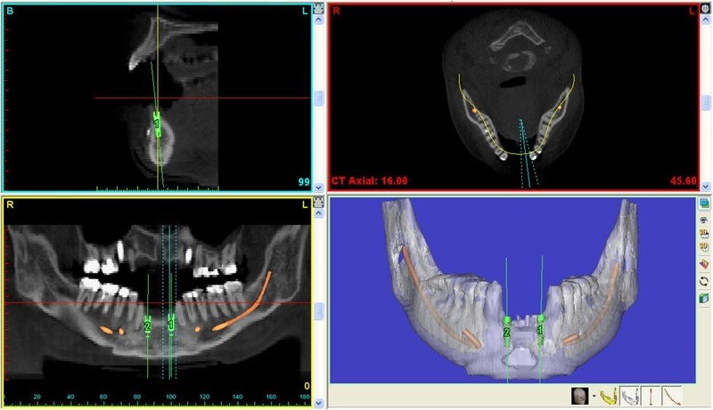
Advances in computer software technology have enabled digitized information from computerized tomography (CT) scans to be used for implant placement planning. The clinician can view and interact with the CT scan data to presurgically place the implant body and visualize the prosthodontic implications.
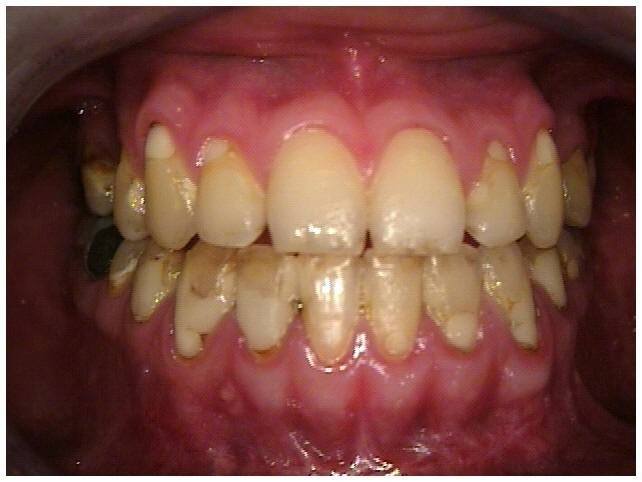
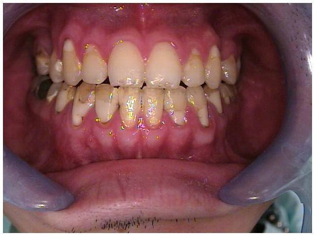
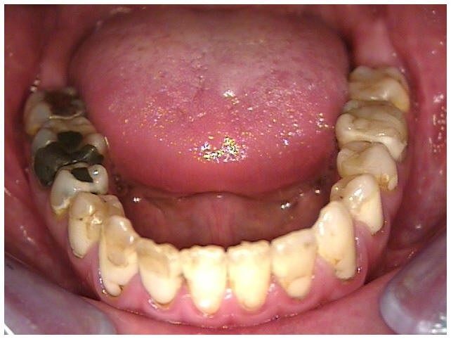
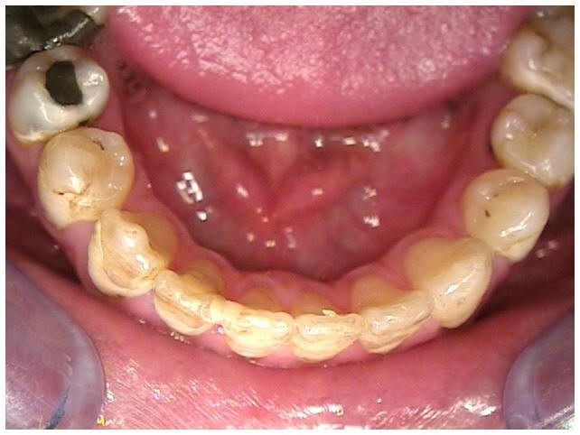
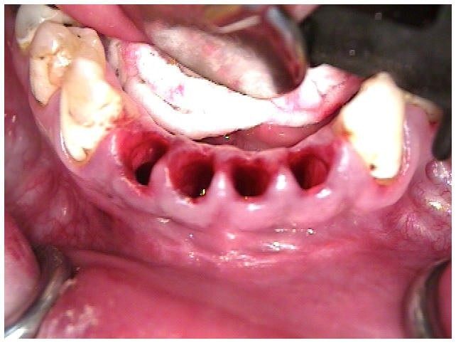
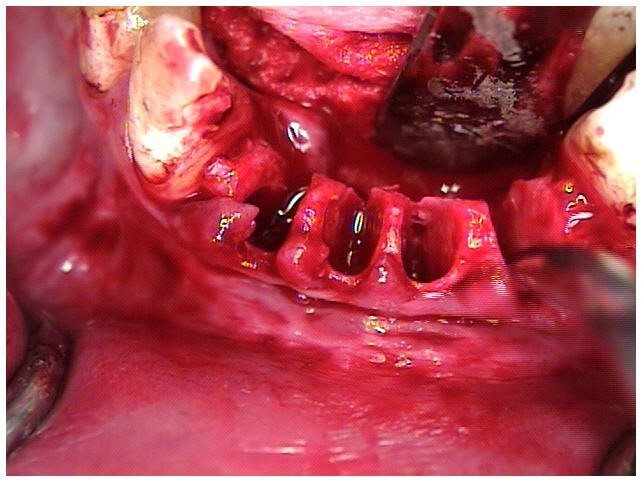
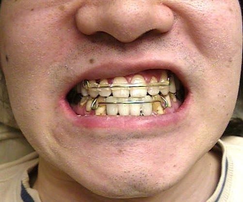
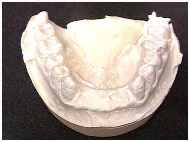
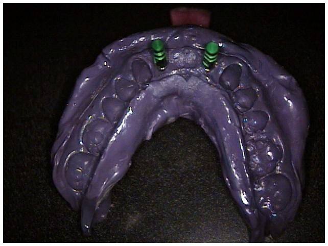
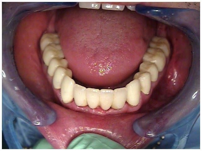
The clinical evaluation of the partially edentulous patient is based on visual examination, manual palpation, gauging tissue thickness and evaluation of soft tissue quality. An appropriate radiographic assessment and accurate pretreatment mounted diagnostic casts are also necessary. During the diagnostic phase of implant treatment, a treatment prosthesis is fabricated for the proposed surgical site following conventional prosthodontic protocol. Patient acceptance of the prosthesis will aid in determining if a fixed implant prosthesis is indicated and if grafting is required to change the quantity of bone present to create the fixed type of prosthesis. The aesthetic and functional positions of the teeth should be determined and accepted by the patient before any radiographic or surgical intervention using the individualized patient acceptance prosthesis. After evaluation and patient approval, the acceptance prosthesis or a duplicate of the existing prosthesis can be used as a CT scan record base and surgical template.
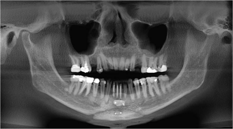
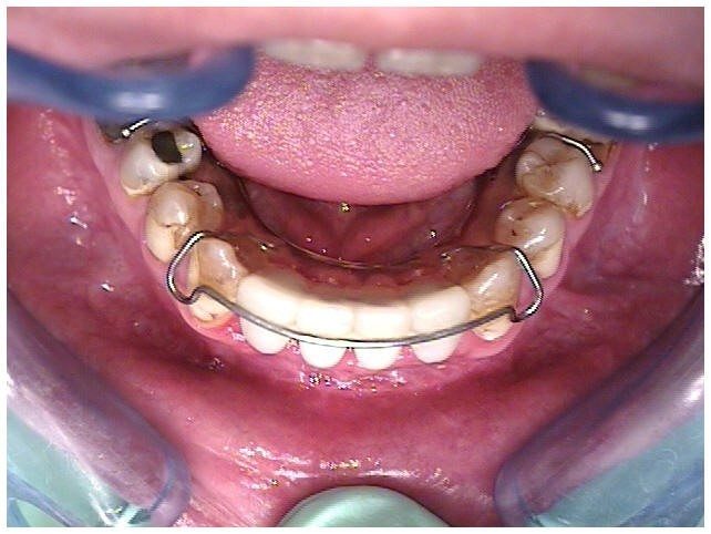
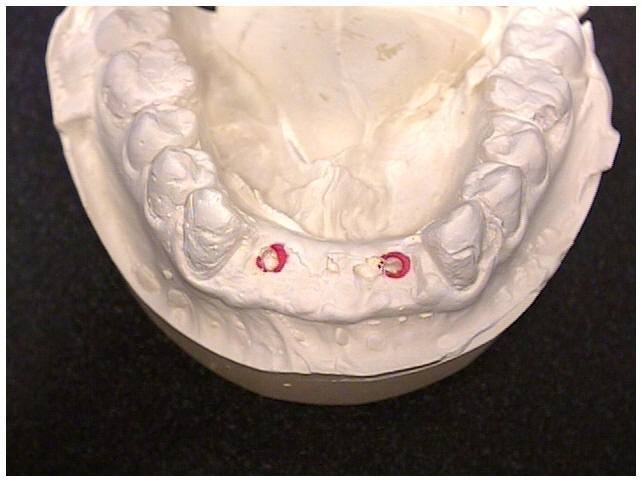
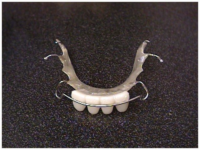
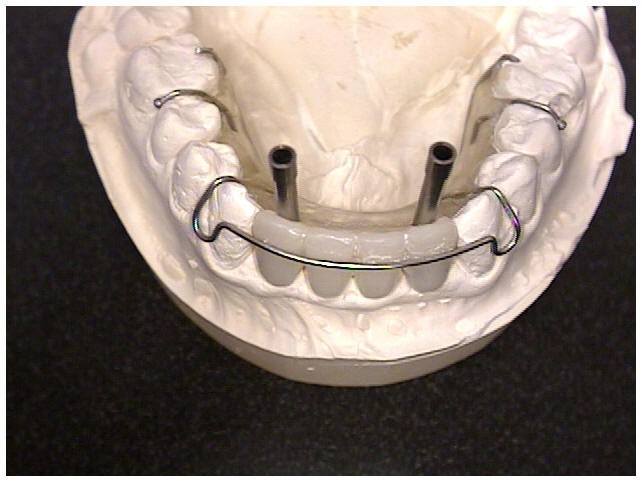
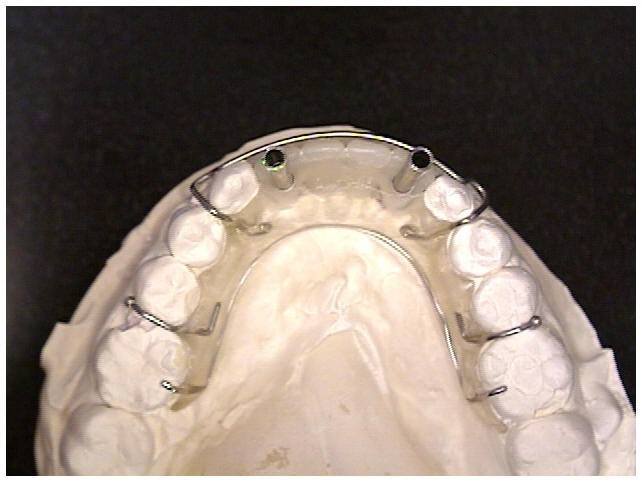
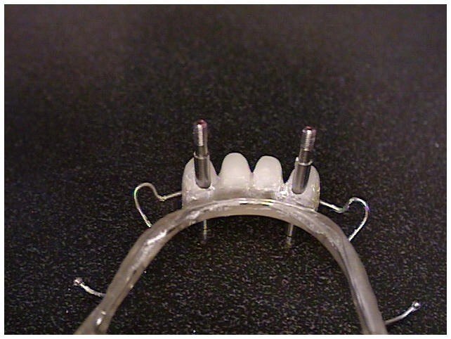
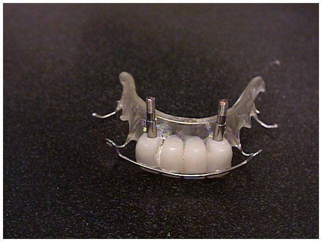
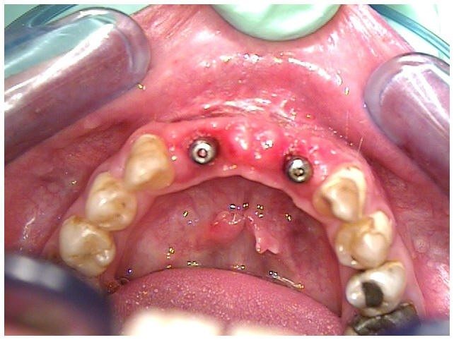
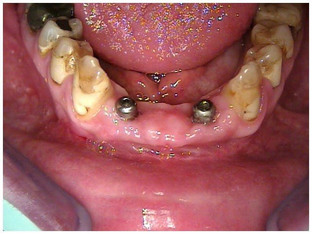
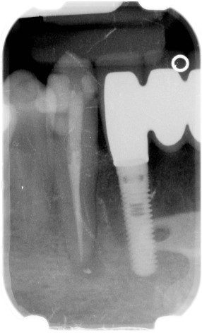
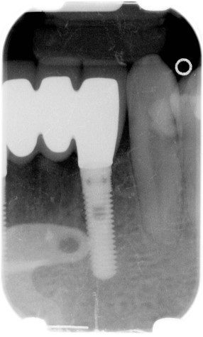
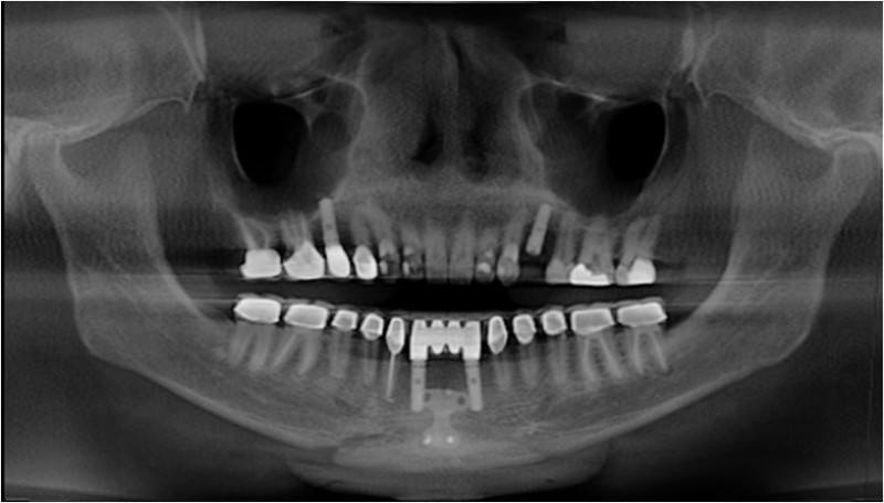
The most frequently used radiograph to survey implant sites is the panoramic view. A panoramic radiograph provides only a two-dimensional view and reveals little of the true, often complex, three-dimensional bony anatomy. It is not uncommon to discover anatomic conditions different from those anticipated based on the limited available information. When implant placement is considered in close proximity to vital structures, a computerized tomographic survey (CBCT Cone Beam Computerized Tomographic Survey) is required for diagnostic and surgical accuracy. The CBCT scan produces a distortion-free, three-dimensional image of the underlying bone that can be further enhanced by the use of a radiopaque CT scan record base. In this patient’s treatment planning, the orthodontic retainer was modified to accept replicas of the natural teeth 23, 24, 25, 26 with contrast media imbedded in the replica teeth for accurate implant position diagnosis with the I-CAT and Simplant studies.
We invite you to contact Reconstructive and Implant Dental Center at 913-534-8801 today to learn more about surgical implant placement in Overland Park, Kansas, and to schedule an appointment with Dr. EDward M. Amet, our prosthodontist.