

The aesthetic restoration of facial appearance and improved dental function with implant prosthodontics is directly related to correctly restoring missing intraoral soft and hard tissues and the aesthetics and technical abilities of the dentist and dental technician. 1 The use of dental implants for oral rehabilitation has revolutionized prosthodontics over the past three decades. Multiple studies have proven the efficacy and excellent long-term prognosis with dental implants.2-5
While initial research and clinical use were directed primarily toward the edentulous patient, more recent studies have focused on the aesthetic and functional use of implants in the partially edentulous patient.6 The most challenging area of modern implant dentistry remains the “aesthetic zone” in the anterior maxilla and mandible. Replacing multiple anterior teeth in the otherwise dentate patient requires careful consideration of the location and volume of residual bone, soft tissue esthetics and room for the implants and prosthesis.
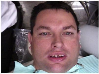
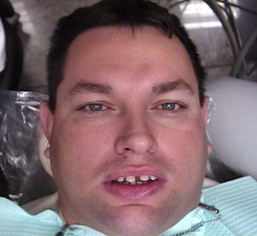
Most dental implants have been and are placed in a delayed manner after tooth extraction allowing for hard and soft tissues to heal prior to implantation. Unfortunately, this allows for resorption of the alveolar ridge in both the buccolingual and coronoapical directions. Studies have shown that as much as 3 to 4 mm of resorption can occur during the first six months post extraction without the intervention of tissue grafting or strengthening techniques.7,8 Dental implants, if placed with a delayed surgical protocol for an implant supported ceramometal reconstruction with only residual native bone, may have unnatural tooth length with spaces between the root structures.
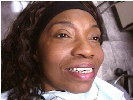
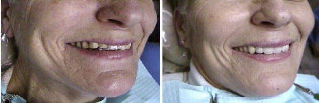
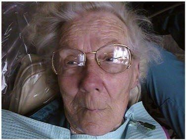
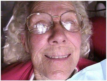
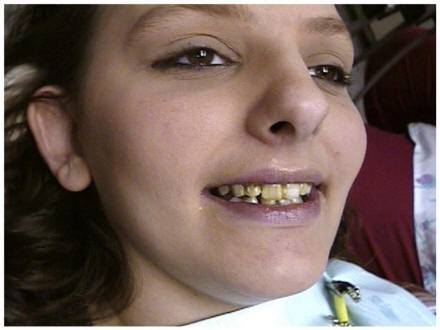
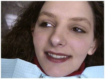
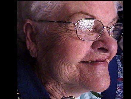
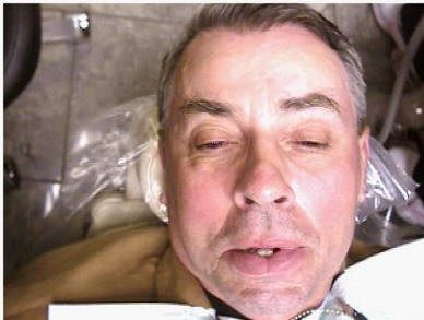
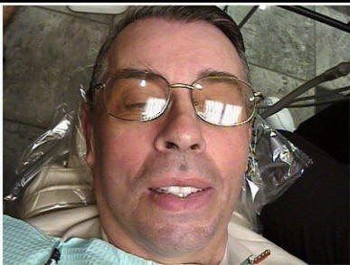
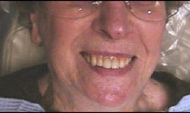
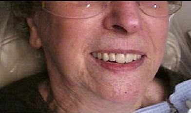
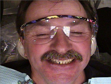
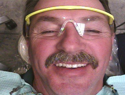
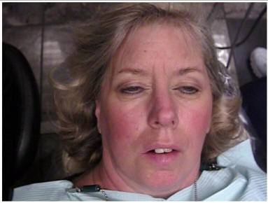
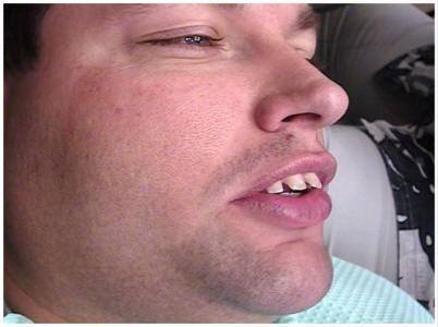
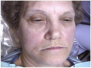
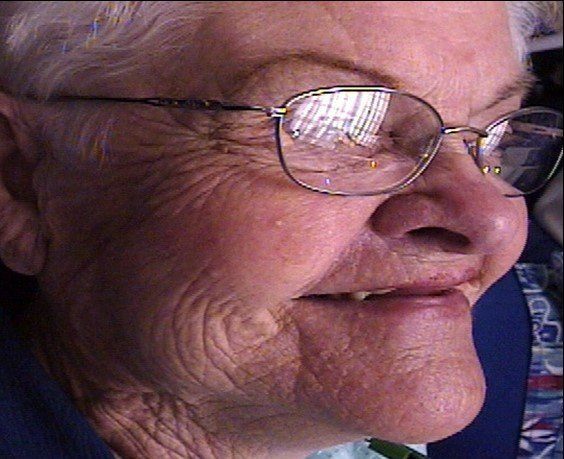
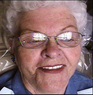
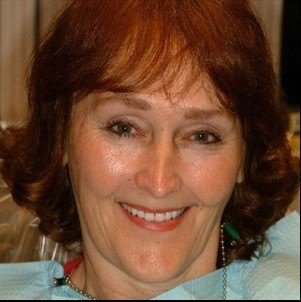
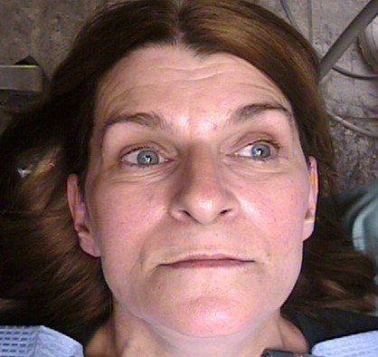
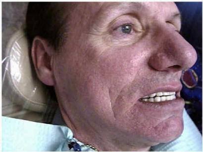
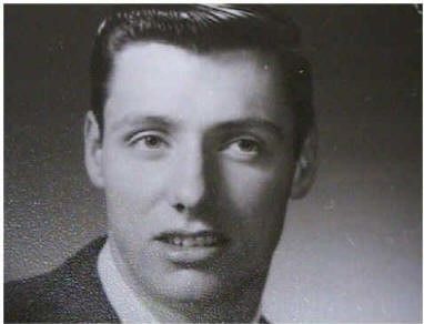
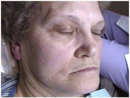
Since facial appearance and normal speech depends on where the teeth are positioned, as well as their shape, form, surface texture and color, it is often not difficult on casual meeting to detect a person who has an implant supported overdenture or fixed partial denture. To compensate for the appearance of the artificial teeth, smaller, shorter teeth are often used and positioned for less visibility. The evenly set smaller teeth can detract from realism and interfere with speech patterns. The lips will often appear lengthened, tense or thin in an attempt to conceal these teeth which may also be set too far posterior in the mouth. The result is the appearance of premature aging which is caused not by age itself but by the change of facial appearance from the chin and nose appearing too close together with the soft tissue compensating for this change in occlusal vertical dimension and decreased facial height. The result is the lack of an aesthetic dental smile with premature aging and may result in the incorrect pronunciation of words with the “f” and “v” sounds.
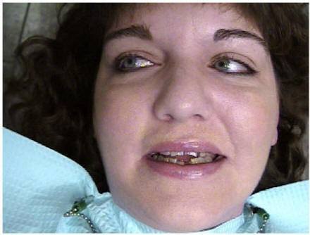
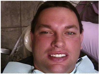
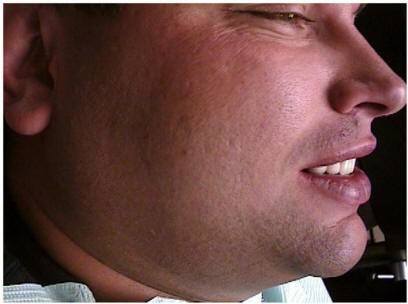
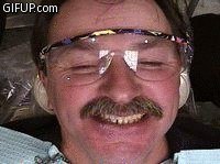
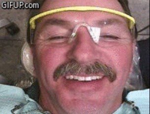
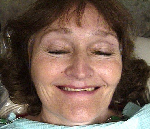
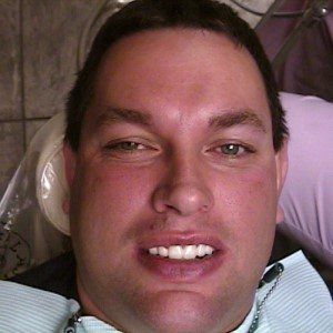
From a dental point of view for correct tooth position, the “s” sound is important to use. This is the case because the “s” sound when articulated is mainly influenced by the teeth and palatal part of the maxillary prosthesis. The sibilants or (sharp sounds) “s”, “z”, “sh”, “ch”, and “j” are alveolar sounds made when the tongue and alveolus form the controlling valve. The important observation when teeth are being replaced is to evaluate the “s” or sibilant sound as it is produced and the relationship of the anterior teeth to each other. The upper and lower incisors should approach end-to-end when the “s” sound is made but should not touch, and this can be recorded digitally during speech production with the use of an extraoral camera at the actual moment the sound is made. This incisal edge sound position is true for class I, II, and III jaw relationship patients. This then allows for the photographic verification of the maxillary and mandibular incisal edge position as the teeth exactly approach end-to-end and indicates horizontal overlap of the anterior teeth, the limits of the current physiologic rest position, and can be used as a measurement for occlusal vertical dimension. This photographic verification will reveal the error but will not indicate whether it is the upper or lower teeth that are incorrectly positioned. After studying the photographs of “s” sound production on the computer monitor and the tooth set-up on the articulator in the profile view, the necessary corrections can be easily seen and thus made to the tooth set-up to allow a physiological aesthetic tooth set-up for either fixed or removable prosthodontics.
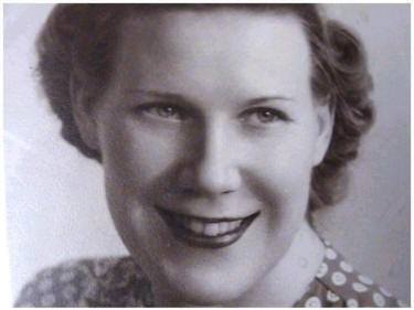
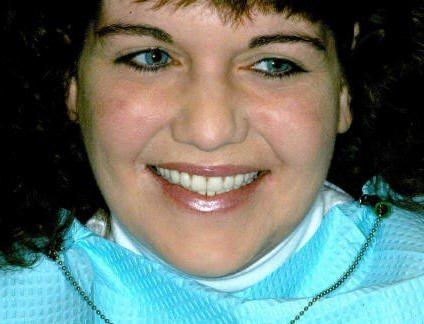
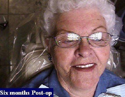
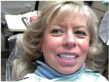
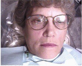
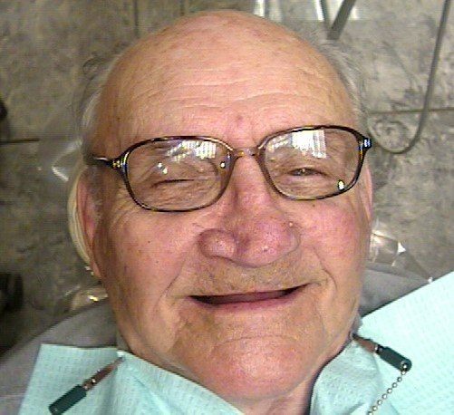
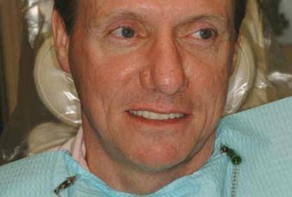
An endosteal implant in bone stimulates and maintains bone dimension and density in a manner similar to healthy natural teeth. As a result of implant stability and prosthetic position, the patient’s facial features are complimented by support with the prosthesis. A totally implant-supported restoration can be positioned for aesthetics, function, and speech, rather than in the “neutral zones” of soft tissue support as in a complete tissue-supported removable prosthesis.
REFERENCES
1. Zarb G, Bolender C, Carlsson, G, Boucher’s Prosthodontic Treatment for Edentulous Patients, Eleventh Edition. St. Louis, The C. V. Mosby Company; 1997.
2. Adell R, Lekholm U, Rockler B, Branemark P-I. A 15-year study of osseointegrated implants in the treatment of edentulous jaw. Int J Oral Surg 1981;10(6):387-416.
3. Branemark P-I., Zarb GA, Alberktsson T (eds). Tissue-Integrated Prostheses. Osseointegration in Clinical Dentistry. Carol Stream, Il; Quintessence, 1985.Linquist LW,
4. Carlsson GE, Glantz PO. Rehabilation of the edentulous mandible with a tissue- integrated fixed prothesis: A 6-year longitudinal study. Quintessence Int 1-987; 18:89-96
5. Laney WR, Tolman D, Keller EE, Desjard RP, Van Roekel NB, Branemark P-I. Dental implants; Tissue-integrated prosthesis utilizing the osseointegration concept. Mayo Clin Proc 1986;61(2):91-97.
6. Chiche GJ, Block MS, Pinault A. Implant surgical template for partially edentulous patients. Int J Oral Maxillofac Implants 1-989-428-9292.
7. Atwood DA, Coyt DA. Clinical, cephalometric and densitometric study of reduction of residual ridges. J Prosthet Dent 1-971-262-80293
8. Johnson K. A study of the dimensional changes occurring in the maxilla after tooth extraction. Part I: Normal healing. Aust Dent J 1-963-842-8433.

Personalized Cosmetic Dentistry Improving and Softening Facial Contours