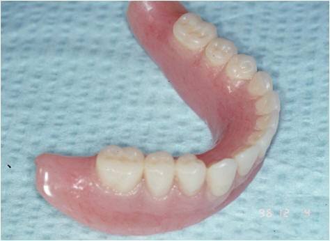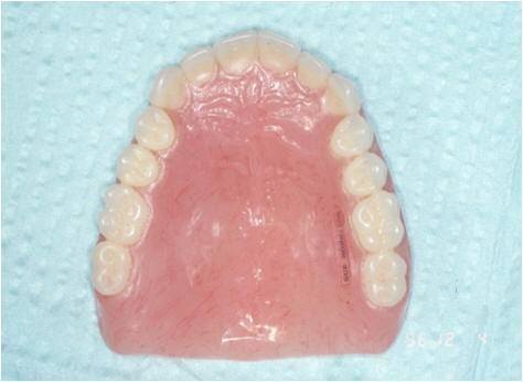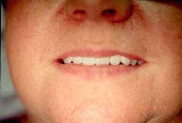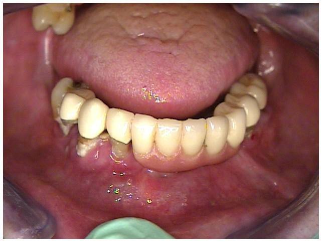
The entire lower half of the face depends on where the teeth are positioned, as well as their shape, form, surface texture and color. It is usually not difficult on casual meeting to detect a person who has artificially constructed teeth or completed dentures. Small, evenly positioned denture teeth detract from realism. The upper lip often appears lengthened and tense or wrinkled in an attempt to conceal the anterior teeth which may be set too far posterior in the mouth. The result is the appearance of premature aging, which is caused not by age itself but by the change of facial appearance from the chin and nose seeming too close together and the soft tissue compensating for this incorrect distance between these parts of the face. The result is the lack of an aesthetic denture smile with premature aging.
The goal of modern dentistry is to achieve integration of dental implants and prosthetic treatment, which replaces a missing tooth or teeth and lost supporting structures, as well as to restore proper aesthetics and function. In order to achieve this goal, it is necessary for the patient and dentist to be able to visualize the final prosthetic results, prior to surgical treatment. Therefore, it is essential for the dentist to have a thorough understanding of all phases of treatment in order to diagnose, treatment plan and restore natural appearance.
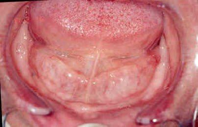
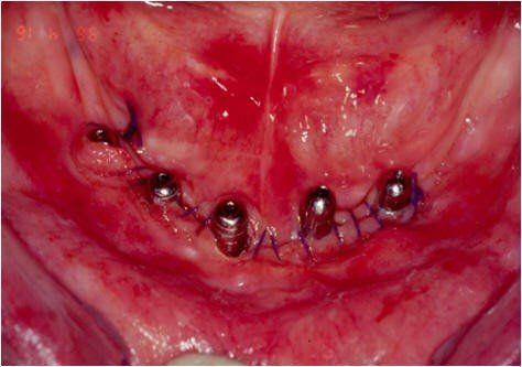
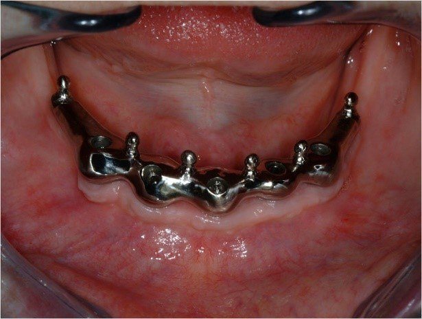
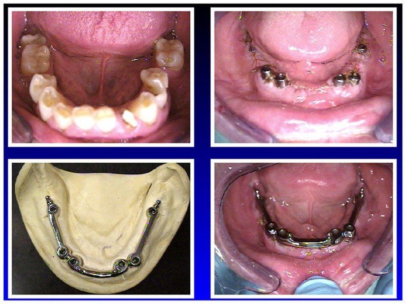
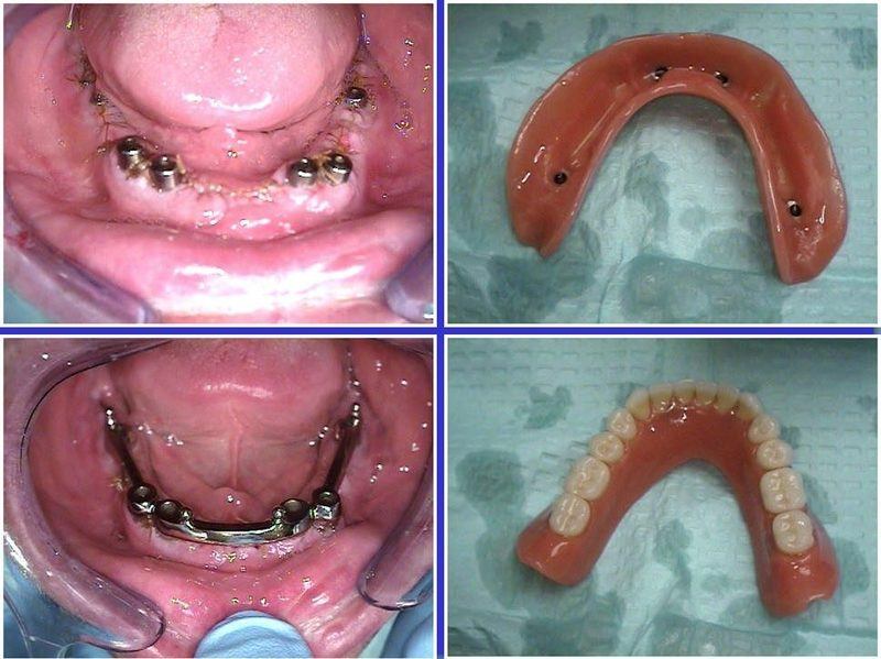
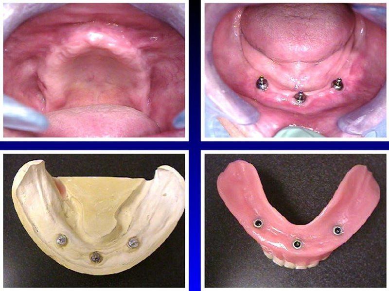
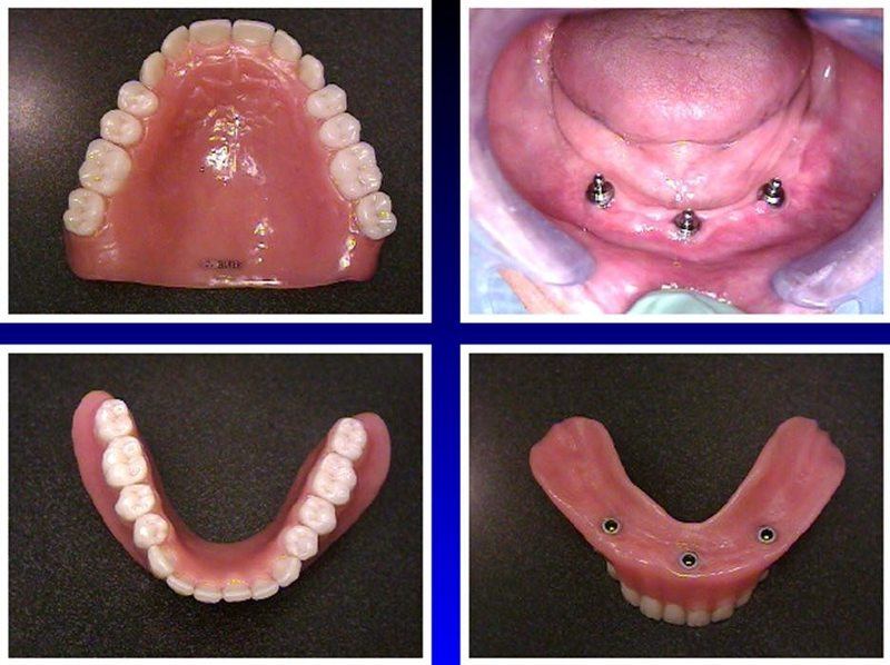
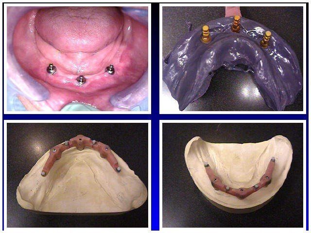
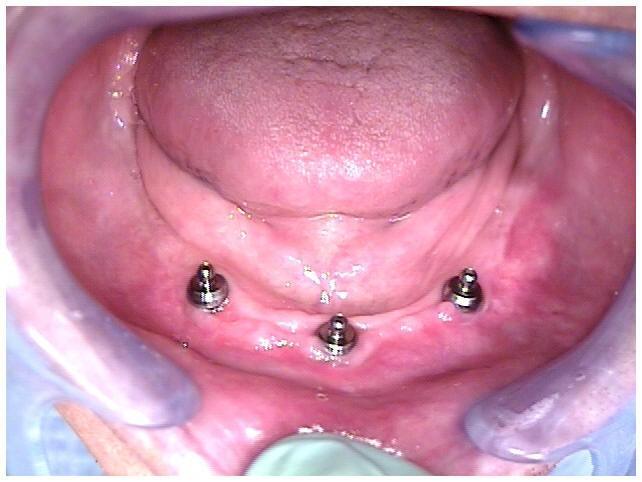
The clinical evaluation of the totally edentulous patients is based on an implant examination with evaluation of soft tissue quality. Appropriate X-rays are needed, as are accurate pre-treatment mounted diagnostic casts. Likewise, during the diagnostic phase of implant treatment planning conventional prosthodontic protocol is followed.
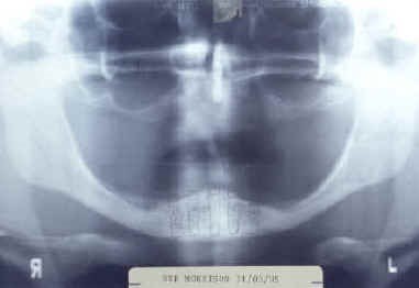
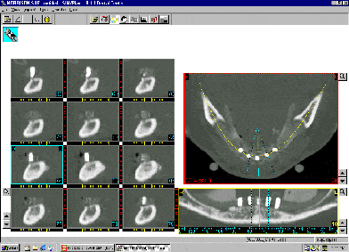
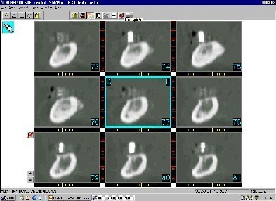
The most frequent X-ray utilized to survey implant sites is the dental panoramic view. This provides a two-dimensional view and reveals little of the true, often complex, three-dimensional bony anatomy. It is not uncommon to discover anatomical conditions different from those anticipated, based on the limited information available. When implant placement is considered in close proximity to vital structures, a computerized tomographic survey (CT scan) is required for diagnostic and surgical accuracy. The CT scan delivers a distortion-free, three-dimensional image of the underlying bone that can be further enhanced by the use of a radiopaque patient acceptance prosthesis or CT scan record base.
Software technology advances (Simplant, Columbia Scientific, Inc., Columbia, MD) permit the CT Scan digital information to be able to be used on desktop or notebook personal computer (PC). All three radiographic views can be viewed simultaneously or individually magnified, and permit the clinician to view and interact with the CT scan data on the computer monitor. It is also possible presurgically to represent the proposed implants, grafting sites, and prosthetic abutments there by avoiding potential prosthetic problems.
The patient acceptance prosthesis is modified with radiopaque markers to make a CT scan record base and utilization in conjunction with the CT scan, facilitates the determination of the prosthetic treatment plan. The prosthetic considerations will dictate the final implant position, if grafting or ridge augmentation is needed and the prosthetic treatment.
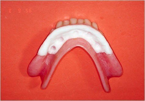
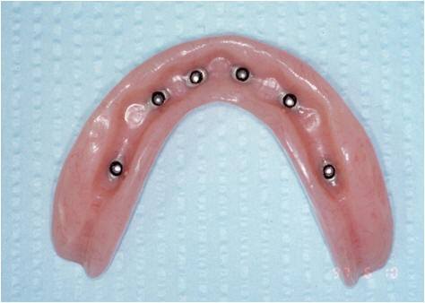
Since the dental laboratory plays such an important role in treatment, having an in-office laboratory is desirable to achieve the final aesthetic and functional result. Finally, the artistic skill of the dentist and technician determines the end result.
We invite you to contact Reconstructive and Implant Dental Center at 913-534-8801 today to learn more about implant-supported bar overdentures in Overland Park, Kansas, and to schedule a visit with our prosthodontist, Dr. EDward M. Amet.